Back Of Neck Anatomy / 1 / Each nerve provides sensation to a specific area of the body called a dermatome.. The superficial lymph nodes of the head and neck receive lymph from the scalp, face and neck. These two ligaments connect and support the spine from the neck to the lower. The spinal cord travels from the base of the skull. It also includes some facts regarding pathophysiology in this region. Anatomy of pelvic bones 12 photos of the anatomy of pelvic bones anatomy bones pelvic region, anatomy pelvic area bones, anatomy pelvic bone female, anatomy pelvic bones and hips, ct anatomy of pelvic bones, human anatomy, anatomy bones pelvic region, anatomy pelvic area bones, anatomy pelvic bone female, anatomy.
The occipital bone surrounds a large opening known as the foramen magnum. The occipital bone is the only bone in your head that connects with your cervical spine (neck). These two ligaments connect and support the spine from the neck to the lower. Extending from underneath the chin, to the posterior aspect of the head. Browse 3,096 anatomy of neck and shoulder stock photos and images available, or start a new search to explore more stock photos and images.
4 353 Neck Anatomy Photos Free Royalty Free Stock Photos From Dreamstime from thumbs.dreamstime.com One of the most important jobs of the cervical spine is to protect the spinal cord as it travels through the neck to innervate the rest of the body. An area called the occiput. The cervical spine supports the weight and movement of your head and protects the nerves exiting your brain. The skull is a strong, bony capsule that rests on the neck and encloses the brain. Browse 3,096 anatomy of neck and shoulder stock photos and images available, or start a new search to explore more stock photos and images. 3d interactive tutorials on the anatomy of the neck, including the anatomical organisation, musculature, larynx, pharynx, blood supply and innervation. The top of the cervical spine connects to the skull, and the bottom connects to the upper back at about shoulder level. It also includes some facts regarding pathophysiology in this region.
The neck is connected to the upper back through a series of seven vertebral segments.
Back of neck anatomy : The lymph nodes and other lymphoid tissues in the head and neck are often swollen and create inflammations, which are encountered by posterior triangle or spinal accessory nodes. Dummies helps everyone be more knowledgeable and confident in applying what they know. They move the head in every direction, pulling the skull and jaw towards the shoulders, spine, and scapula. The neck is the area between the skull base and the clavicles. Below the neck, holding the tooth into the bone, is the root of the tooth. The neurocranium (cranial vault) and the viscerocranium (facial skeleton). It is made up of bones, discs, muscles, ligaments, nerves and tendons. Neck muscles can be strained from poor posture — whether it's leaning over your computer or hunching over your workbench. The neck is connected to the upper back through a series of seven vertebral segments. The occipital bone surrounds a large opening known as the foramen magnum. These muscles are mainly responsible for the movement of the head in all directions. Cervical spine anatomy video the cervical spine has 7 stacked bones called vertebrae, labeled c1 through c7.
These muscles give the sides of the neck their. Anatomy of pelvic bones 12 photos of the anatomy of pelvic bones anatomy bones pelvic region, anatomy pelvic area bones, anatomy pelvic bone female, anatomy pelvic bones and hips, ct anatomy of pelvic bones, human anatomy, anatomy bones pelvic region, anatomy pelvic area bones, anatomy pelvic bone female, anatomy. They move the head in every direction, pulling the skull and jaw towards the shoulders, spine, and scapula. It consists of two major parts: See human neck anatomy stock video clips.
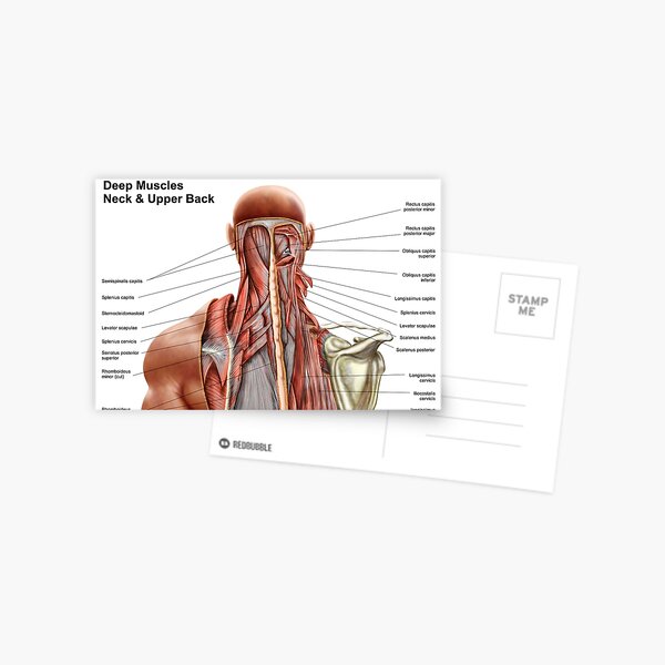
Human Anatomy Showing Deep Muscles In The Neck And Upper Back Postcard By Stocktrekimages Redbubble from ih1.redbubble.net The cervical spine supports the weight and movement of your head and protects the nerves exiting your brain. The muscles of the neck are present in four main groups. They are arranged in a ring shape; It is made up of bones, discs, muscles, ligaments, nerves and tendons. Cervical spine anatomy video the cervical spine has 7 stacked bones called vertebrae, labeled c1 through c7. The top of the cervical spine connects to the skull, and the bottom connects to the upper back at about shoulder level. They consist of 3 main groups of muscles: Below the neck, holding the tooth into the bone, is the root of the tooth.
Each nerve provides sensation to a specific area of the body called a dermatome.
Rarely, neck pain can be a symptom of a more serious problem. The neurocranium (cranial vault) and the viscerocranium (facial skeleton). Back of neck anatomy : It is made up of bones, discs, muscles, ligaments, nerves and tendons. The occipital bone is a bone that covers the back of your head; It also includes some facts regarding pathophysiology in this region. 3d video tutorials and interactive modules on the anatomy of the back including anatomy of the musculature, vertebral column, joints and ligaments. They ultimately drain into the deep lymph nodes. Extending from underneath the chin, to the posterior aspect of the head. The lymph nodes and other lymphoid tissues in the head and neck are often swollen and create inflammations, which are encountered by posterior triangle or spinal accessory nodes. Anatomy of back of neck. They move the head in every direction, pulling the skull and jaw towards the shoulders, spine, and scapula. These muscles give the sides of the neck their.
This entry was posted in anatomy by admin. The skull is a strong, bony capsule that rests on the neck and encloses the brain. The larynx is located where the pharynx, the back of the mouth and nasal cavity, divides into the trachea (the tube that carries air to the lungs) and the esophagus (the tube that carries food to. Browse 3,096 anatomy of neck and shoulder stock photos and images available, or start a new search to explore more stock photos and images. 3d interactive tutorials on the anatomy of the neck, including the anatomical organisation, musculature, larynx, pharynx, blood supply and innervation.
Anatomy Of The Back Spine And Back Muscles Kenhub from thumbor.kenhub.com Anterior, lateral and posterior groups, based on their position in the neck. It also includes some facts regarding pathophysiology in this region. The suboccipital muscles act to rotate the head and extend the neck.rectus capitis posterior major and rectus capitis posterior minor attach the inferior nuchal line of the occiput to the c2 and c1 vertebrae respectively.obliquus capitis superior also extends from the occiput to c1 while obliquus capitis inferior originates from c2 and. Back of neck region anatomy : This entry was posted in anatomy by admin. The neck muscles, including the sternocleidomastoid and the trapezius, are responsible for the gross motor movement in the muscular system of the head and neck. Spinal cord anatomy in the neck. The occipital bone is the only bone in your head that connects with your cervical spine (neck).
Pain in a man's body pain in a man's body on a gray background.
Anterior, lateral and posterior groups, based on their position in the neck. See more ideas about back pain, spine health, spine problems. Rarely, neck pain can be a symptom of a more serious problem. The spinal cord is a bundle of nerves that carry electrical signals between the brain and the rest of the body. The occipital bone is the only bone in your head that connects with your cervical spine (neck). These muscles give the sides of the neck their. It also includes some facts regarding pathophysiology in this region. Below the neck, holding the tooth into the bone, is the root of the tooth. The neck is connected to the upper back through a series of seven vertebral segments. The muscles of the neck are muscles that cover the area of the neck . Delphian node) lie between the back of neck anatomy. The suboccipital muscles act to rotate the head and extend the neck.rectus capitis posterior major and rectus capitis posterior minor attach the inferior nuchal line of the occiput to the c2 and c1 vertebrae respectively.obliquus capitis superior also extends from the occiput to c1 while obliquus capitis inferior originates from c2 and. Each nerve provides sensation to a specific area of the body called a dermatome.